
What does a bacterial flange actually look like? Going way beyond the microscope, researchers from across several schools at USC and The Scripps Research Institute in La Jolla, Calif., gathered this month to discuss the latest advances in how scientists can accurately capture images of the tiniest particles and live biological processes.
The jam-packed daylong retreat at USC featured presentations from more than a dozen scientists, who discussed a variety of approaches to imaging from four-dimensional modeling to the challenges of electron microscopy. The researchers’ work ranges from better imaging of nanoparticles to body organs to capturing a realistic range of emotions in the human face.
“Scripps and USC created this retreat because of our mutual interest in the convergence of technology and molecular science, working to understand the structure of life,” said Randolph Hall, vice president of research at USC, who organized the event with researchers in the fields of biology, medicine, cinema, lighting and engineering.
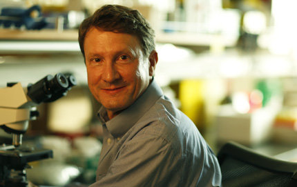
Among USC’s presenters was Scott Fraser, a world leader in using advanced imaging techniques to capture biological processes and Provost Professor of Biological Sciences and Biomedical Engineering and the Director of Science Initiatives at USC Dornsife and USC Viterbi School of Engineering. Fraser’s colorful images of living tissues imaged to the cellular level stunned the group.
One challenge of microscopy is that the light required to create images can also damage living cells—prompting those on the cutting edge of microscopy to generate ever faster ways of capturing images. Fraser said his goal is to image living organisms—which, in his case, are typically zebrafish—“from the embryo to them crawling out of the microscope without losing any cells.”
Other USC presenters included:
• Andrew McMahon, director of the Eli and Edythe Broad Center for Regenerative Medicine and Stem Cell Research, who discussed imaging of the kidney, including the use of fluorescent genes and different patterns of branching among kidney cells;
• Paul Debevec, associate director for graphics research at the USC Institute for Creative Technologies, whose lighting techniques have been used to create visual effects for films such as Gravity and Avatar;
• Richard Weinberg of the USC School of Cinematic Arts, a pioneer in filming and live-streaming microscopic subjects, whose digital movies of microorganisms include In the Pond, featuring the life teeming in pond water;
• Noah Malmstadt of the USC Viterbi School of Engineering, who demonstrated lipid bilayer deformation and disruption by cationic nanoparticles;
• Justin Haldar of USC Viterbi, who discussed the challenges of signal-to-noise in the reconstruction of images, including the possibility of setting known constraints.
USC researchers were joined on the retreat by faculty from the Scripps Research Institute, who discussed ongoing work including methods of improving and speeding up the process of electron microscopy and ways to visualize the actual motion of the tiny molecular machines that allow cells to function.
“We are fortunate to have Scripps as our partner,” Hall said. “Together we can reveal the mechanisms underlying human development, disease and evolution.”