Skip to content

Menu
  • USC Stem Cell
  • About
    • Stem Cell FAQs
    • Mission and History
    • California’s Leadership in Stem Cell Research
    • Founding Supporters and Ambassadors
    • Well-being
    • Jobs
  • News & Events
    • News
    • Events
    • Videos
    • Impact Reports and Newsletters
  • Research
    • Department Faculty
    • Eli and Edythe Broad Center Faculty
    • Research Facilities
    • USC+CHLA Alpha Clinic
    • Translational Research Committee
    • USC Stem Cell Research Oversight Committee (SCRO)
    • Apply to Become Center Faculty
  • Education & Training
    • Undergraduate
    • Master’s Program
    • PhD Program
    • Medical Education
    • Postdoctoral Opportunities
    • Our Trainees
  • Funding
  • Inclusive Excellence
  • Support Us
  • Contact
    • Directory
    • Subscribe
  • Search

USC Stem Cell study breaks the silence on how fish and lizards regenerate hearing

By  Cristy Lytal

Posted December 9, 2024
Reading Time 3 minutes

in this section

  • News
  • Events
  • Videos
  • Impact Reports and Newsletters

read this next

The organ of Corti, the hearing organ of the inner ear, is made up of a single row of inner hair cells and three rows of outer hair cells (green), surrounded by supporting cells (purple). (Image by Yassan Abdolazimi and Neil Segil)

USC Stem Cell researchers listen for clues about how the gene Atoh1 enables hearing

  • Follow us on
  • Like us on
  • Follow us on
  • Follow us on
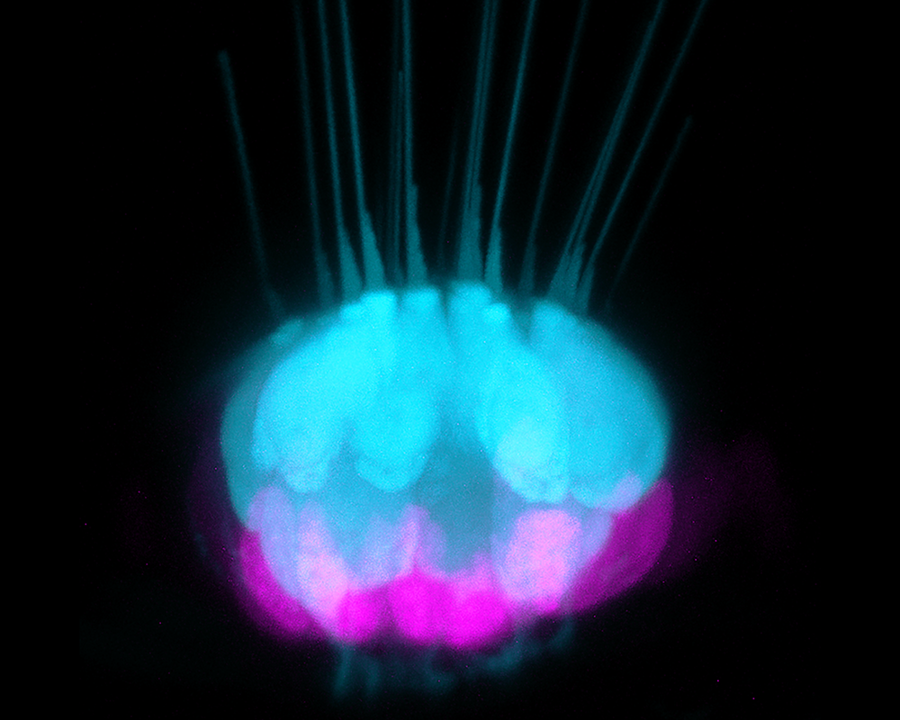
In the zebrafish inner ear, a cell type known as supporting cells (magenta) gives rise to new sensory hearing cells (blue). The study reveals a set of DNA control elements critical for supporting cells to regenerate hair cells after injury in zebrafish, lizards and other regenerative species. (Image by Tuo Shi/Crump and Lozito labs/USC Stem Cell)

A new USC Stem Cell study published in the Proceedings of the National Academy of Sciences (PNAS) has identified key gene regulators that enable some deafened animals—including fish and lizards—to naturally regenerate their hearing. The findings could guide future efforts to stimulate the regeneration of sensory hearing cells in patients with hearing loss and balance disorders.

Led by first author Tuo Shi and co-corresponding authors Ksenia Gnedeva and Gage Crump at the Keck School of Medicine of USC, the study focuses on two cell types in the inner ear: the sensory cells that detect sound, and the supporting cells that create an environment where sensory cells can thrive. In highly regenerative species such as fish and lizards, supporting cells can also transform into replacement sensory cells after injury—a capacity absent in humans, mice and all other mammals.

To better understand this remarkable regenerative process, the scientists determined how genes normally only found in the sensory cells can be re-activated in the supporting cells of regenerative species. To achieve that, the scientists determined how the genome is folded in the sensory cells and supporting cells of the inner ears of regenerative zebrafish and green anole lizards. They then compared DNA control elements for sensory genes in zebrafish and green anole lizards to those in mice, which cannot replace sensory hearing cells after injury.

“By comparing two different regenerative vertebrates—zebrafish and lizards—to non-regenerative vertebrates such as mice, we found something that was fundamental to the way sensory cells can be replaced to restore hearing in some vertebrates,” said Crump, professor of stem cell biology and regenerative medicine at USC.

Their experiments revealed a class of DNA control elements known as “enhancers” that, after injury, amplify the production of a protein called ATOH1, which in turn induces a suite of genes required to make sensory cells of the inner ear.

Using CRISPR, a gene editing tool, the scientists deleted five of these enhancers in zebrafish, impairing both the formation of sensory hearing cells during development and their regeneration following injury.

“In the past, deletion of individual enhancers most often does not have much of an effect,” said Crump. “But by targeting all five enhancers in zebrafish, we discovered their critical role in both development and regeneration.”

Interestingly, although zebrafish also possess the same type of sensory cell in a specialized aquatic organ called the lateral line, which senses water flow and pressure, the genetic deletions only impacted cells in their inner ears.

The researchers found that mice possess equivalent enhancers that are active during embryonic development in the progenitor cells that give rise to the sensory and supporting cells of the inner ear. However, only regenerative species such as fish and lizards maintain these enhancers in an open configuration in their supporting cells into adulthood, preserving their capacity to replace damaged sensory cells.

“What we have found is that sister cell types in regenerative vertebrates maintain open enhancers from development into adult stages, thus allowing these related cells to replace each other following damage,” said Crump. “In the future, targeted strategies to open up these enhancers in the human inner ear could be used to boost our natural regenerative abilities and reverse deafness.”

Additional authors include Yeeun Kim, Juan Llamas, Xizi Wang, Peter Fabian, Thomas P. Lozito, and the late Neil Segil from USC.

This work was funded by the National Institute on Deafness and Other Communication Disorders (grants F31DC020633, T32DC009975, and R01DC020268), the National Institute of General Medical Sciences (grant R01GM115444), and the Hearing Restoration Project funded by the Hearing Health Foundation.

Read more about: Brain Nerves and Senses
Mentioned in this article: Gage Crump, PhD, Ksenia Gnedeva, PhD, Thomas Lozito, PhD

Post navigation

← To remember conversations, keep making new brain cells
USC selected to receive $3.4 million award from ARPA-H’s Sprint for Women’s Health →
Keck School of Medicine of USC
1975 Zonal Ave.
Los Angeles, CA 90033
Google Map
Phone: (323) 442-1900
Hours:
Monday–Friday
7:30am–5:00pm PST
Resources For
  • Current Students
  • Faculty & Staff
  • Patients
  • Community
  • Press
  • Visitors
Areas of Focus
  • Education & Training
  • Research
  • Patient Care
  • Community
Departments and Offices
  • Departments
  • Institutes and Centers
  • Research Programs
  • Administrative Offices
About Keck
  • History
  • Leadership
  • Annual Report
  • Digital Accessibility
Intranet
  • Privacy Notice
  • Notice of Non-Discrimination
  • Smoke-Free Policy

Copyright © 2025 University of Southern California

  • Research
    • Research HomeCutting-edge research drives innovation in healthcare at the Keck School of Medicine
    • Where Research Happens
    • Research Funding
    • Training and Education
    • Researcher Resources
    • Collaborate and Partner
  • Education
    • Education HomeNurturing future healthcare leaders through excellence in education
    • MD Program
    • Residencies and Clinical Fellowships
    • PhD Programs
    • Master’s Programs
    • Professional Programs
    • Post-Doctoral Researchers
    • CME, Certificate & Undergraduate Programs
  • Departments, Institutes & Centers
    • Basic Science and Clinical DepartmentsExploring foundational science and specialized clinical fields
    • Institutes and Centers
    • Research Programs
  • About
    • About the Keck SchoolDiscover the mission, history, and vision of the Keck School of Medicine
    • History
    • Leadership
    • Dean’s Corner
    • Life in Southern California
    • Visit
  • Our Faculty
  • Current Students
  • Newsroom
  • Events Calendar
  • Support the Keck School
  • USC.edu
  • Are you a Patient?