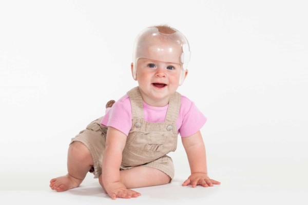
USC scientists have regenerated parts of the skull affected by a common birth defect called craniosynostosis.
Using stem cells to regenerate parts of the skull, USC scientists partially corrected a skull deformity and reversed learning and memory deficits in young mice with craniosynostosis, a condition estimated to affect 1 in every 2,500 infants born in the United States.
The only current therapy is complex surgery within the first year of life, but skull defects often return afterward. The study, which appears today in Cell, could pave the way for more effective and less invasive therapies for children with craniosynostosis.
“I started my career as a clinician treating kids with congenital defects, and we always wanted to do something better for these patients,” said study leader Yang Chai, a University Professor and director of the Center for Craniofacial Molecular Biology at the Herman Ostrow School of Dentistry of USC.
“This stem cell-mediated suture regeneration approach truly gives us the hope that one day we can apply this as a biological solution for this biological problem.”
Healthy infants are born with soft spots on their heads called sutures, which are comprised of flexible tissue that fills the space between the skull bones. Sutures allow the skull to expand as the brain grows rapidly in the first few years of life.
In craniosynostosis, one or more sutures fuse and turn into bone too early, closing the gap between skull plates, increasing pressure inside the skull and leading to abnormal growth. The increased pressure in the skull causes physical changes in the brain that lead to thinking and learning problems.
“We wanted to know if restoring sutures could improve neurocognitive function in mice genetically engineered with Twist1 mutation, which is known to cause craniosynostosis in humans,” said Chai, who is also the associate dean of research at the Ostrow School.
Replenishing stem cells could be “life-changing” for kids with craniosynostosis
The scientists focused on a group of stem cells found in sutures called Gli1+ cells. Previous studies by the group indicated that the cells are key to keeping skull sutures of young mice intact. The team also found that Gli1+ cells are depleted just before skull bones prematurely fuse in mice with craniosynostosis. The scientists reasoned that replenishing the cells might regenerate the flexible sutures in affected animals.
To test this idea, the researchers took Gli1+ cells from healthy mice, suspended the cells in a biodegradable gel and placed the mixture in grooves where the fused skull sutures used to be in mice with craniosynostosis.
Six months after surgery, skull imaging and tissue analysis showed that new fibrous, flexible sutures had formed in treated areas. The regenerated sutures remained intact even a year after surgery. In contrast, the same grooves closed in mice that received a gel that lacked Gli1+ cells.
Untreated mice with craniosynostosis had increased pressure inside their skulls and poor performance on tests of social and spatial memory and motor learning. After treatment, most of these measures all returned to levels typical of healthy mice. The skull deformities of treated mice were also partially reversed.
The treatment also reversed the loss of brain volume and nerve cells in areas involved in learning and memory. According to the scientists, these findings shed light on the mechanisms underlying impaired brain function and its improvement after suture regeneration.
The scientists note that more work remains before such an intervention can be tested in humans, including studies to figure out the optimal timing of surgery and the ideal source and amount of stem cells.
“Currently, these operations involve moving the bones of the forehead and skull while operating on top of the brain. To be able to avoid those risks would obviously be a tremendous paradigm shift for the field of craniofacial surgery,” said Mark Urata, a craniofacial surgeon at the Keck School of Medicine of USC and a co-author of the study.
“This could be life-changing for so many babies yet to be born.”
In addition to Chai and Urata, Jianfu Chen of the Ostrow School and postdoctoral fellow Li Ma performed all the neurocognitive studies. Other authors on the study include Mengfei Yu, Yuan Yuan, Jinzhi He and Thach-Vu Ho of the Center for Craniofacial Molecular Biology at USC; Axel Montagne, Yingxi Wu, Zhen Zhao, Naoma Sta Maria, Russell Jacobs and Berislav V. Zlokovic of the Zilkha Neurogenetic Institute at USC; and Xin Ye and Huiming Wang of the Key Laboratory of Oral Biomedical Research at the Zhejiang University School of Medicine in Hangzhou in China.
This research was supported by NIDCR grants DE026339, DE012711, DE022503 and DE026914. Support also came from the National Institute of Neurological Disorders and Strokes grant NS097231.
