
In a proof-of-concept study, a research team based at the Keck School of Medicine of USC showed that functional ultrasound imaging can record brain activity through a transparent skull implant.
In the first study of its kind, researchers from the Keck School of Medicine of USC and the California Institute of Technology (Caltech) designed and implanted a transparent window in the skull of a patient, then used functional ultrasound imaging (fUSI) to collect high-resolution brain imaging data through the window. Their preliminary findings suggest that this sensitive, non-invasive approach could open new avenues for patient monitoring and clinical research, as well as broader studies of how the brain functions.
“This is the first time anyone had applied functional ultrasound imaging through a skull replacement in an awake, behaving human performing a task,” said Charles Liu, MD, PhD, a professor of clinical neurological surgery, urology and surgery at the Keck School of Medicine and director of the USC Neurorestoration Center. “The ability to extract this type of information noninvasively through a window is pretty significant, particularly since many of the patients who require skull repair have or will develop neurological disabilities. In addition, ‘windows’ can be surgically implanted in patients with intact skulls if functional information can help with diagnosis and treatment.”
The research participant, 39-year-old Jared Hager, sustained a traumatic brain injury (TBI) from a skateboarding accident in 2019. During emergency surgery, half of Hager’s skull was removed to relieve pressure on his brain, leaving part of his brain covered only with skin and connective tissue. Because of the pandemic, he had to wait more than two years to have his skull restored with a prosthesis.
During that time, Hager volunteered for earlier research conducted by Liu, Jonathan Russin, MD, associate surgical director of the USC Neurorestoration Center, and another Caltech team on a new type of brain imaging called fPACT. The experimental technique had been done on soft tissue, but could only be tested on the brain in patients like Hager who were missing a part of their skull. When the time came for implanting the prosthesis, Hager again volunteered to team up with Liu and his colleagues, who designed a custom skull implant to study the utility of fUSI—which cannot be done through the skull or a traditional implant—while repairing Hager’s injury.
Before the reconstructive surgery, the research team tested and optimized fUSI parameters for brain imaging, using both a phantom (a scientific device designed to test medical imaging equipment) and animal models. They then collected fUSI data from Hager while he completed several tasks, both before his surgery and after the clear implant was installed, finding that the window offered an effective way to measure brain activity. The research, funded in part by the National Institutes of Health, was just published in the journal Science Translational Medicine.
Functional brain imaging, which collects data on brain activity by measuring changes in blood flow or electrical impulses, can offer key insights about how the brain works, both in healthy people and those with neurological conditions. But current methods, such as functional magnetic resonance imaging (fMRI) and intracranial electroencephalography (EEG) leave many questions unanswered. Challenges include low resolution, a lack of portability or the need for invasive brain surgery. fUSI may eventually offer a sensitive and precise alternative.
“If we can extract functional information through a patient’s skull implant, that could allow us to provide treatment more safely and proactively,” including to TBI patients who suffer from epilepsy, dementia, or psychiatric problems, Liu said.
A new frontier for brain imaging
As a foundation for the present study, Liu has collaborated for years with Mikhail Shapiro, PhD and Richard Andersen, PhD, of Caltech, to develop specialized ultrasound sequences that can measure brain function, as well as to optimize brain-computer interface technology, which transcribes signals from the brain to operate an external device.
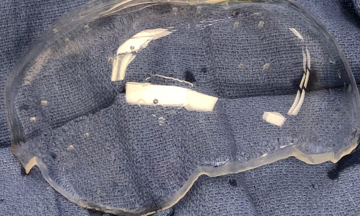
With these pieces in place, Liu and his colleagues tested several transparent skull implants on rats, finding that a thin window made from polymethyl methacrylate (PMMA)—which resembles plexiglass—yielded the clearest imaging results. They then collaborated with a neurotechnology company, Longeviti Neuro Solutions, to build a custom implant for Hager.
Before surgery, the researchers collected fUSI data while Hager did two activities: solving a “connect-the-dots” puzzle on a computer monitor and playing melodies on his guitar. After the implant was installed, they collected data on the same tasks, then compared the results to determine whether fUSI could provide accurate and useful imaging data.
“The fidelity of course decreased, but importantly, our research showed that it’s still high enough to be useful,” Liu said. “And unlike other brain-computer interface platforms, which require electrodes to be implanted in the brain, this has far less barriers to adoption.”
fUSI may offer finer resolution than fMRI and unlike intracranial EEG, it does not require electrodes to be implanted inside the brain. It is also less expensive than those methods and could provide some clinical advantages for patients over non-transparent skull implants, said Russin, who is also an associate professor of neurological surgery at the Keck School of Medicine and director of cerebrovascular surgery at Keck Hospital of USC.
“One of the big problems when we do these surgeries is that a blood clot can form underneath the implant, but having a clear window gives us an easy way to monitor that,” he said.
Refining functional ultrasound technology
In addition to better monitoring of patients, the new technique could offer population-level insights about TBI and other neurological conditions. It could also allow scientists to collect data on the healthy brain and learn more about how it controls cognitive, sensory, motor and autonomic functions.
“What our findings shows is that we can extract useful functional information with this method,” Liu said. “The next step is: What specific functional information do we want, and what can we use it for?”
Until the new technologies undergo clinical trials, fUSI and the clear implant are experimental. In the meantime, the research team is working to improve their fUSI protocols to further enhance image resolution. Future research should also build on this early proof-of-concept study by testing more participants to better establish the link between fUSI data and specific brain functions, the researchers said.
“Jared is an amazing guy,” said Liu, who is continuing to collaborate with the study participant on refining new technologies, including laser spectroscopy, which measures blood flow in the brain. “His contributions have really helped us explore new frontiers that we hope can ultimately help many other patients.”
About this research
In addition to Liu, Russin, Shapiro and Andersen, the study’s other authors are Kay Jann, PhD, from the Mark and Mary Stevens Neuroimaging and Informatics Institute, Keck School of Medicine of USC; Claire Rabut, Sumner Norman and Whitney Griggs from the California Institute of Technology; and Vasileios Christopoulos from the University of California Riverside.
This work was supported by the National Institutes of Health [R01NS123663]; the T&C Chen Brain-Machine Interface Center; the Boswell Foundation; the National Eye Institute [F30 EY032799]; the Josephine de Karman Fellowship; the UCLA-Caltech Medical Scientist Training Program [NIGMS T32 GM008042]; the Della Martin Postdoctoral Fellowship; the Human Frontier Science Program Cross-Disciplinary Fellowship [LT000217/2020-C]; the USC Neurorestoration Center; and the Howard Hughes Medical Institute.
Disclosure
Claire Rabut, Whitney S. Griggs, Sumner L. Norman, Richard A. Andersen, Charles Liu and Mikhail G. Shapiro have filed a provisional patent application based on this research.