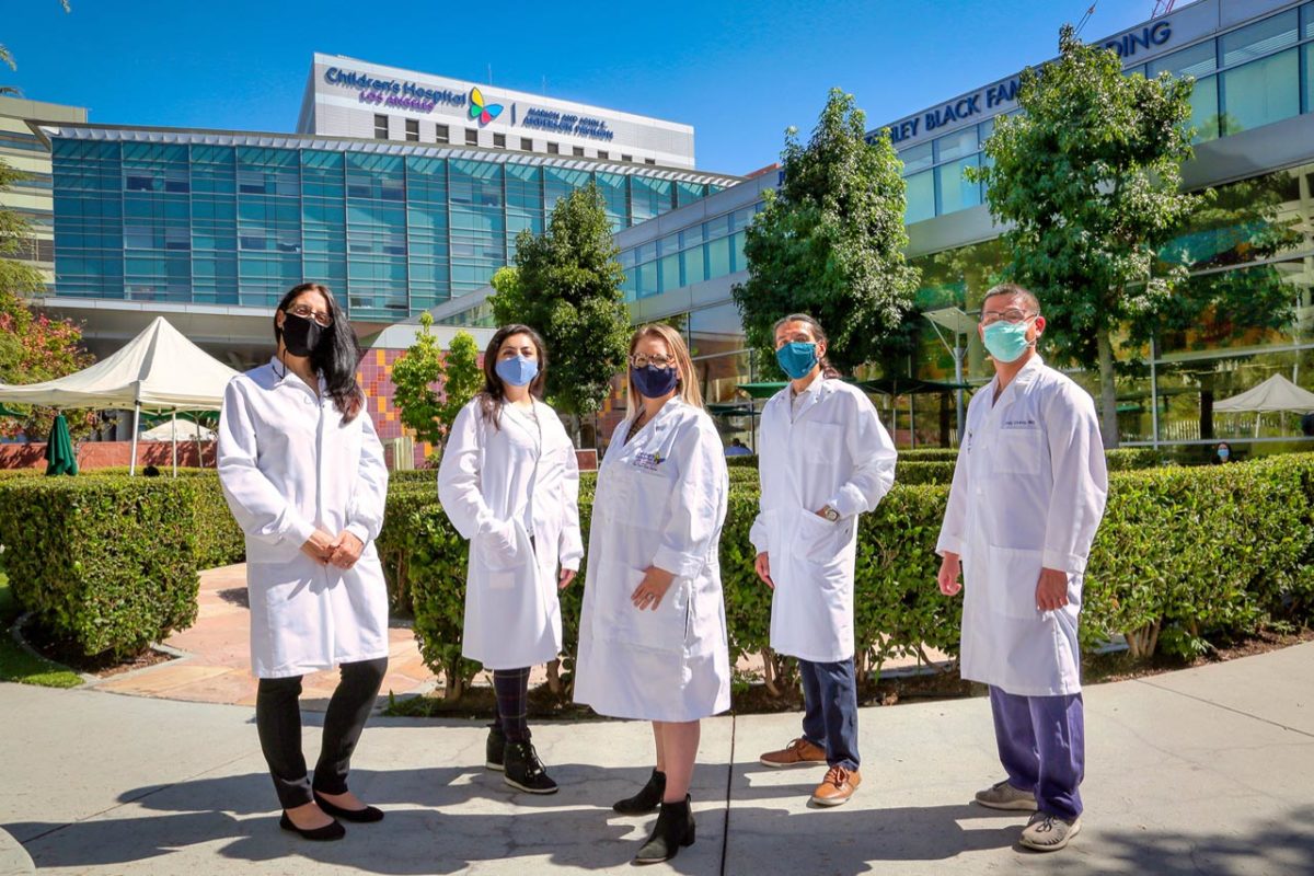
Stefano Da Sacco, PhD, calls Wilms tumor “an underdog” in the research world.
“If you go to the American Urological Association or other meetings, there aren’t many presentations or discussions about Wilms tumor,” Dr. Da Sacco says. “From my experience, it’s understudied. Not many basic scientists are working on it.”
It’s a different story at Children’s Hospital Los Angeles. Laura Perin, PhD, and Dr. Da Sacco are not only studying Wilms tumor—they’re at the forefront of research in the field. They also work closely with the clinicians in the Wilms tumor program in the Division of Urology.
“Having that relationship with a basic science lab helps us as surgeons better understand the biology of Wilms tumor and refine our surgeries,” says Andy Chang, MD, Director of Urologic Oncology and Vice Chief of the Division of Urology at Children’s Hospital Los Angeles. “It really primes us for translational approaches.”
Why does Wilms tumor grow?
The most common kidney cancer in children, Wilms tumor has survival rates of over 90%. But about 15% of patients suffer a relapse after treatment and have much lower survival rates.
Children’s Hospital Los Angeles is a major West Coast referral center for Wilms tumor. The multidisciplinary program—which includes pediatric urologists, oncologists, radiation oncologists and pediatric surgeons—has extensive expertise in caring for children with the disease. That includes those with large tumors, bilateral tumors or tumors with unfavorable histology.
But the most unique aspect is the program’s close connection to the GOFARR Laboratory for Organ Regenerative Research and Cell Therapeutics in Urology, part of The Saban Research Institute of Children’s Hospital Los Angeles. The lab is co-led by Roger De Filippo, MD, Chief of the Division of Urology, and Dr. Perin.
Dr. Perin and Dr. Da Sacco—along with Senior Postdoctoral Fellow Astgik Petrosyan, PhD—are studying the molecular mechanisms that regulate the growth of Wilms tumors, as well as those that distinguish between favorable and unfavorable tumors.
The goal is to find a more precise way to classify tumors at diagnosis—and then develop novel therapies that can specifically target those tumor types.
The team, which in 2019 received a grant from The Pablove Foundation, was one of the few groups in the world to isolate live nephron progenitor cells from Wilms tumor tissue samples and transplant them into preclinical models.
Wilms tumor results from uncontrolled proliferation of nephron progenitors—stem cells charged with forming the kidney during development in the womb. Most patients are diagnosed between the ages of 1 and 4; the tumor can be as large as a cantaloupe.
“After birth, these progenitor cells should normally be gone. But in Wilms tumor, they still exist,” Dr. Perin notes. “It’s a development that tries to go on and on, even after birth. The question is, why do the cells do that?”
The team has identified a possible reason: two integrin signaling pathways that may play a key role in keeping the cells in a state where they continually replicate. Through in vivo studies, the researchers are looking at whether blocking those pathways can slow or stop tumor growth.
A bridge from lab to clinic
Collaborating with the clinical team is an important part of their work. Surgeons often share samples of Wilms tumors that have been removed in patients, allowing the lab to conduct more studies. But the partnership also includes robust discussions and exchanges of ideas.
“Knowing what the clinicians need in terms of potential therapies or diagnostics guides the way we look at things,” Dr. Da Sacco adds. “Combining both our points of view helps us move forward faster and better.”
Meanwhile, clinicians are conducting their own research. Dr. Chang, Research Coordinator Zoe Baker, PhD, and Pediatric Urology Fellow Arthi Hannallah, MD, have created a database of 150 Wilms tumor patients and are using it to study factors that contribute to safer partial nephrectomies. Currently, most patients undergo a complete nephrectomy (removal of the affected kidney), although partial removals are done in certain cases.
Dr. Chang’s ultimate hope is that the lab’s studies will one day lead to improved treatments—and allow more children’s kidneys to be saved.
“We’re invested in seeing the best outcomes for children, and by extension, their parents,” he says. “This is a very survivable cancer for most kids. We want to expand that survival to include all kids, while also saving as much of their kidneys as possible.”
