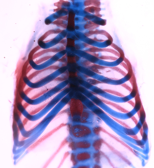
Unlike salamanders, mammals can’t regenerate lost limbs, but they can repair large sections of their ribs.
In a new study in the Journal of Bone and Mineral Research, a team directed by USC Stem Cell researcher Francesca Mariani takes a closer look at rib regeneration in both humans and mice.
The first author of the paper, USC medical student Marissa K. Srour, was a USC undergraduate when she started the project, which earned a 2011 USC Discovery Scholar Prize. Each year, 10 graduating seniors win these coveted prizes, which recognize exceptional new scholarship.
Using CT imaging, Srour, Mariani and their colleague Janice Lee from the University of California, San Francisco, monitored the healing of a human rib that had been partially removed by a surgeon. The eight centimeters of missing bone and one centimeter of missing cartilage did partially repair after six months.
To better understand this repair process, they removed sections of rib cartilage — ranging from three to five millimeters — from a related mammal, mice. When they removed both rib cartilage and its surrounding sheath of tissue — called the “perichondrium,” the missing sections failed to repair even after nine months. However, when they removed rib cartilage but left its perichondrium, the missing sections entirely repaired within one to two months.
They also found that a perichondrium retains the ability to produce cartilage even when disconnected from the rib and displaced into nearby muscle tissue — further suggesting that the perichondrium contains progenitor or stem cells.
“We believe that the development of this model in the mouse is important for making progress in the field of skeletal repair, where an acute clinical need is present for ameliorating skeletal injury, chronic osteoarthritis and the severe problems associated with reconstructive surgery,” said Mariani, assistant professor of Cell and Neurobiology and principal investigator in the Eli and Edythe Broad Center for Regenerative Medicine and Stem Cell Research at USC. “At the early stages in our understanding, the mouse provides us with an exceptional ability to make progress, and we are excited about the potential for using cells derived from the rib perichondrium or using rib perichondrium-like cells for regenerative therapy.”
Additional co-authors include: Jennifer L. Fogel, Kent T. Yamaguchi, Aaron P. Montgomery, Audrey K. Izuhara, Aaron L. Misakian, Stephanie Lam and Daniel L. Lakeland from USC; and Mark M. Urata from Children’s Hospital Los Angeles.
Funding came from an Oral and Maxillofacial Surgery Foundation Research Award; the Baxter Medical Scholar Research Fellowship; USC undergraduate fellowships; the Provost, Dean Joan M. Schaeffer, and Rose Hills fellowships; a California Institute for Regenerative Medicine (CIRM) training fellowship; CIRM BRIDGES fellowships through California State University, Fullerton, and Pasadena City College; and the James H. Zumberge Research and Innovation Fund.
The lab also received support for this study and future work from a new National Institutes of Health (NIH) Exploratory/Developmental Research Grant Award (R21AR064462) — given to high-risk, high-reward studies that break new ground or embark in novel directions. The lab earned this prestigious $450,000 grant from the NIH’s National Institute of Arthritis and Musculoskeletal and Skin Diseases (NIAMS).
The Mariani Lab will also continue its pioneering research through a second new grant from the Merck Investigator Studies Program as well as through a USC Regenerative Medicine Initiative Award with colleagues Gage Crump and Jay Lieberman.
As Mariani explained, “These grants will allow us to address several key questions: Which cells are involved in mediating the repair? How big of a piece of rib can we take out and still see repair? And can we use cells from the rib to get repair in another part of the skeleton? By answering these questions, we are accelerating the discovery of new regenerative therapies for the patients who need them the most.”
